Border Podiatry Centre The “Lateral” Ankle Sprain
Lateral ankle ligament reconstruction is a surgery to tighten and firm up one or more ankle ligaments on the outside of your ankle. It's also known as the Brostrom procedure. It's most often done as an outpatient surgery, so you can go home the same day. Your ankle is a hinge joint that allows motion up and down, and from side to side.
Anterior talofibular ligament tear & sprain, causes, symptoms, diagnosis & treatment
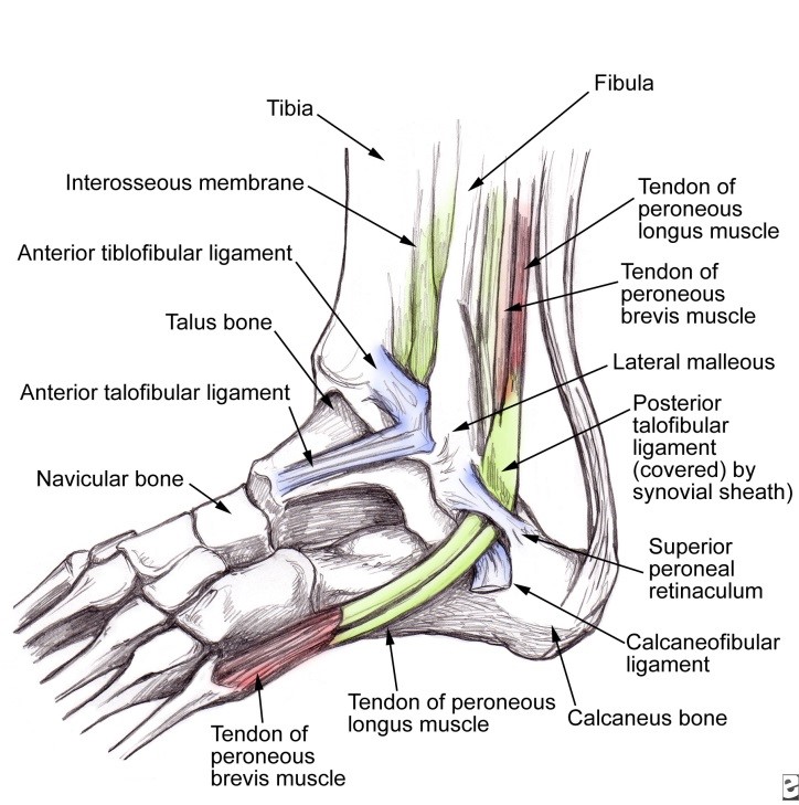
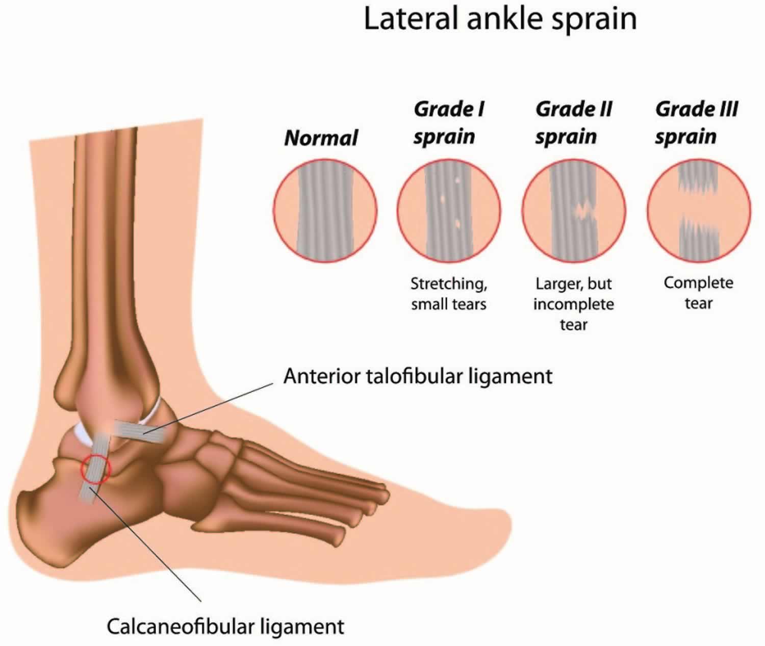
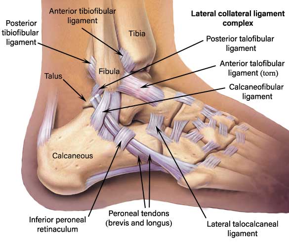
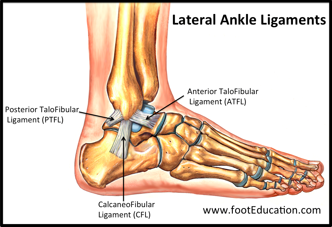
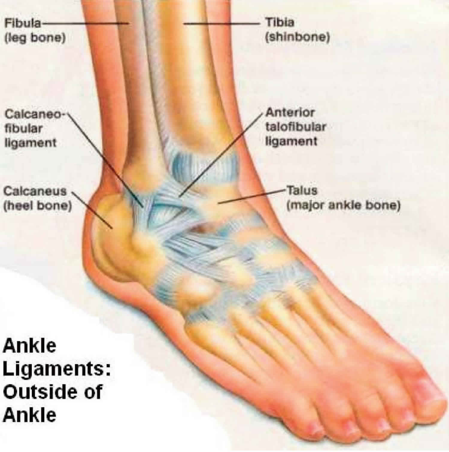
The ligament on the outside of the ankle (lateral ligament) is made up of three separate bands: one at the front (anterior talo-fibular ligament), one in the middle (calcaneo-fibular ligament) and one at the back (posterior talo-fibular ligament). The front and middle bands are the ligaments injured in a sprain.

Torn Ligament in Ankle, Medically 3D Illustration Stock Illustration Illustration of tibia
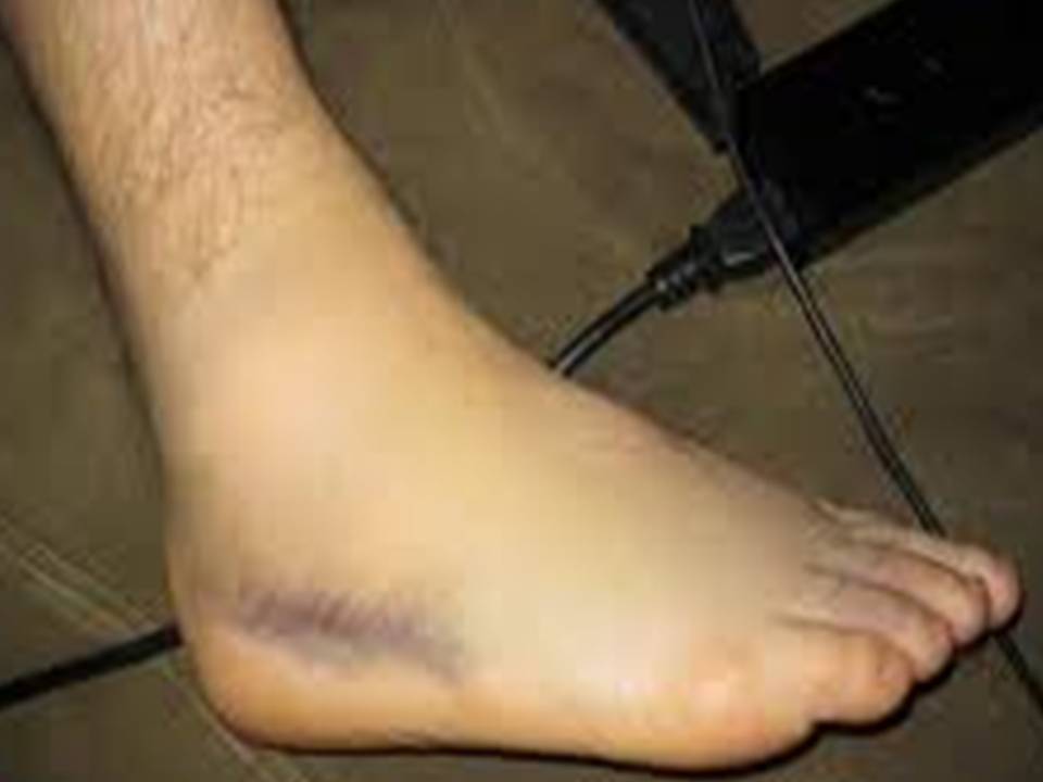
A sprained ankle is an injury to one or more ligaments in the ankle. Mild sprains may involve overstretching and irritating the ligaments, while severe sprains can cause the ligaments to tear.

2 Ligaments Torn In Ankle Lateral Ankle Ligament Injury Physio Check
A sprained ankle is an injury that occurs when you roll, twist or turn your ankle in an awkward way. This can stretch or tear the tough bands of tissue (ligaments) that help hold your ankle bones together. Ligaments help stabilize joints, preventing excessive movement.
Ankle sprains Greg Robinson Podiatrist
Mar 17, 2021 When To Seek Treatment for An Ankle Ligament Tear A good rule of thumb for an ankle sprain is to follow at-home treatment advice for two to three weeks. If the symptoms of pain, swelling and bruising haven't reduced or you experience ongoing instability after the injury, you should get in touch with a specialist.
Ankle Sprain Symptoms, Diagnosis and Treatment MedFog
Magnetic resonance imaging (MRI) is the imaging modality of choice for diagnosing ligament pathologies because of its multiplanar capability and high soft tissue contrast. With MRI, it is possible to triage and attribute the cause of post traumatic ankle pain to bone, ligament, or tendon pathologies, which otherwise overlap clinically.
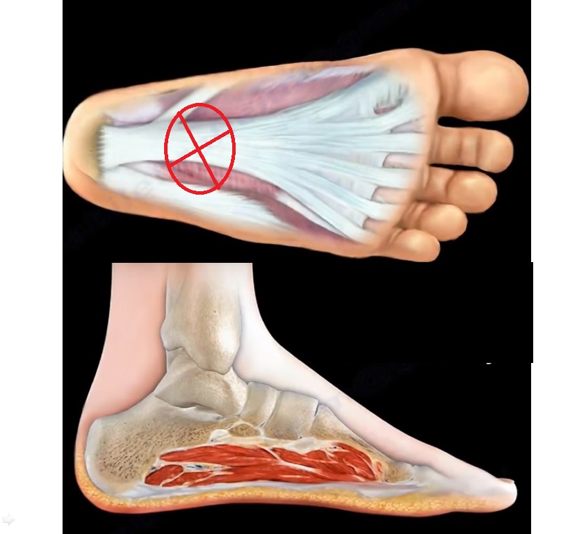
Rupture of the Plantar fascia —
The strong, fibrous ligaments attach bone to bone - and can be torn or stretched as a result of a wrenching movement or an impact. There are four types of ankle sprain: Grade I - stretched ligament or a very mild tear, with little or no instability at the joint. Grade II - more serious but still incomplete tear, with some looseness in the.

Torn Ligament or Foot Tendon
Ligament tears can be surgically repaired by stitching them back together, re-attaching them to the bone, or replacing them with a graft (a tendon from another part of your body or from a donor). Most ankle ligament surgery is carried out using keyhole (arthroscopic) surgery. Mr Simon Moyes is a renowned consultant orthopaedic and sports.
Ankle Fractures Broken Ankle Florida Orthopaedic Institute
Diagnosing torn lateral ankle ligaments is usually through physical examination and x-rays to identify any potential fractures. During the assessment the physician will determine: The degree of instability Loss of strength - Resisted eversion assessment Loss of range of motion (ROM): Dorsiflexion and Plantarflexion - Eversion and Inversion

ankle sprain, ATFL ligament, ankle, pain Sprained ankle, Sprain, Ankle ligaments
Proven risk factors Previous or existing ankle injury especially if poorly rehabilitated (biggest risk factor). Lack of strength and stability related to the ankle. Lack of, or extreme flexibility, in the ankle joint. Poor balance. Sudden change in direction (acceleration or deceleration). Increasing age of player. Suspected risk factors

Torn Ligament in my Ankle HubPages
This article provides a comprehensive review of the normal and injured ankle ligaments on MRI, with emphasis on the anatomy, biomechanics, and imaging features of each ligament. It also discusses the common mechanisms of injury, the classification of ligament tears, and the associated findings of ankle instability. The article is useful for radiologists, orthopedists, and sports medicine.

Torn Ligament in Ankle, 3D Illustration Stock Illustration Illustration of feet, major 182458905
Richard Nilsen Medically Reviewed by Mary D. Daley, MD, MSc Image Credit: skynesher/E+/GettyImages Ligaments are strong, fibrous tissues that hold bones together. Several ligaments around the ankle provide stability to the ankle joint. These ligaments can be stretched or torn when a twisting injury of the ankle occurs.
Sprained Ankle FootEducation
The most common type of ankle sprain is an inversion injury, or lateral ankle sprain. The foot rolls inward, damaging the ligaments of the outer ankle — the anterior talofibular ligament, the calcaneofibular ligament, and the posterior talofibular ligament. (Ligaments are bands of fibrous tissue that connect bone to bone; see illustration.)

Ankle Sprains The Institute for Athletic Medicine
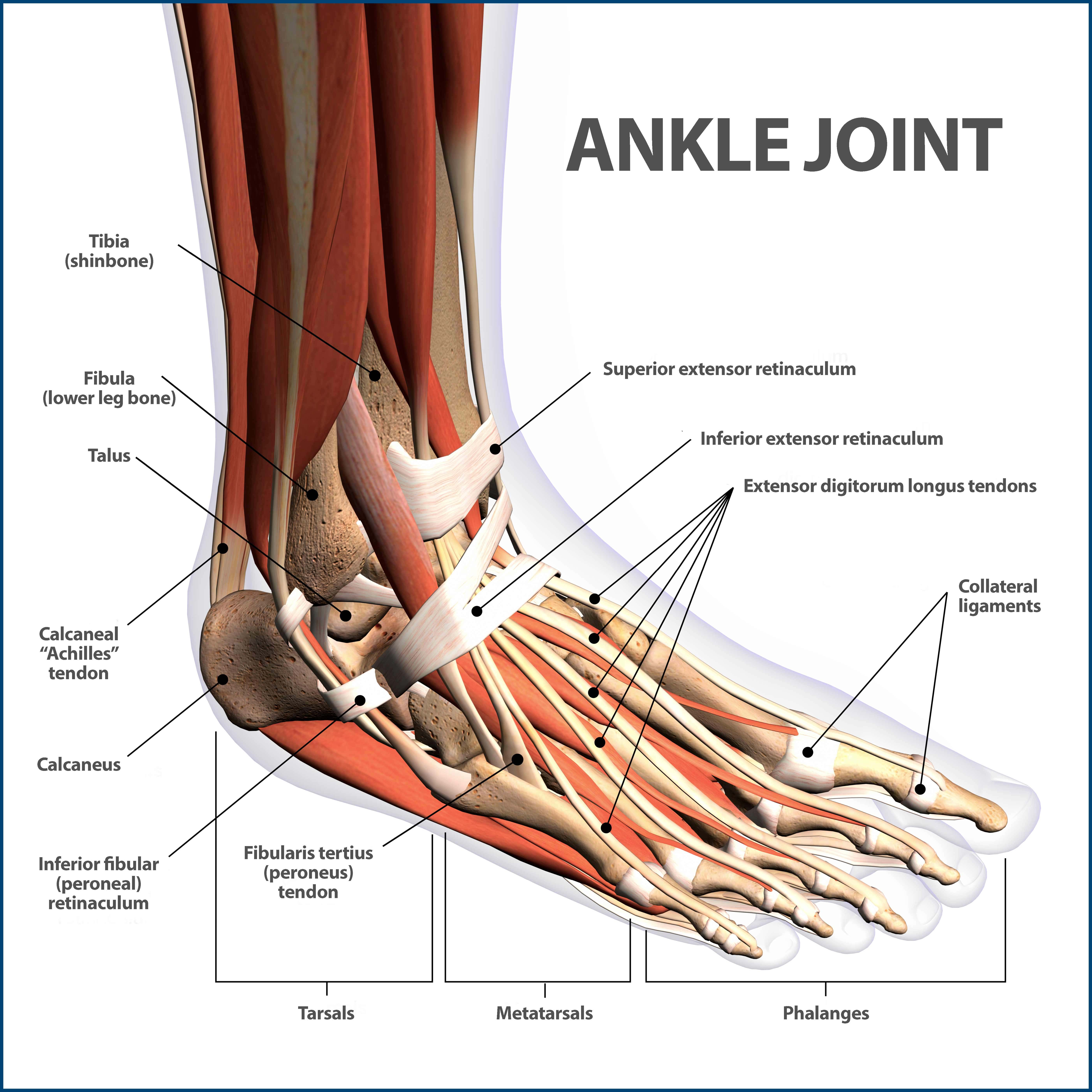
Anatomy What are ankle ligaments made of? Ankle ligaments are made of connective tissue that contains: Collagen, a protein that binds tissues in animals. Slightly stretchy elastic fibers. Advertisement Where are the ankle ligaments located? Ankle ligaments are found throughout your foot, ankle and lower leg. They connect certain bones:
Anterior talofibular ligament tear & sprain, causes, symptoms, diagnosis & treatment
Browse 927 ankle ligaments photos and images available, or start a new search to explore more photos and images. of 16 NEXT United States Browse Getty Images' premium collection of high-quality, authentic Ankle Ligaments stock photos, royalty-free images, and pictures.

Pin on Joints Treatment
1 Images Ankle Joint Osteology Ankle Joint consists of tibial plafond medial malleolus lateral malleolus talus motion main motion plantar flexion dorsiflexion secondary motions inversion/eversion rotation Distal tibiofibular joint consists of distal fibula incisura fibularis concave surface of distal lateral tibia motion